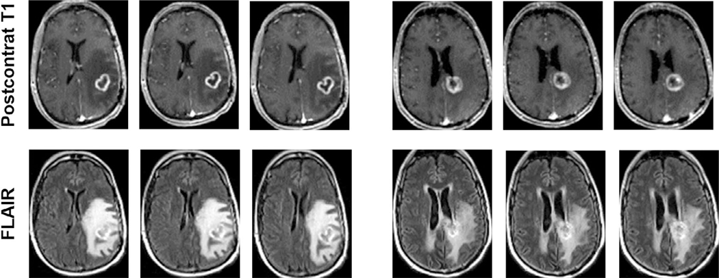
FIGURE 1:
Postcontrast T1-weighted and T2-FLAIR images at three different slice levels from patients with TP (left panel) and PsP (right panel) demonstrating equivocal imaging findings with similar patterns of contrast enhancement and surrounding areas of T2-FLAIR signal abnormality suggesting the limitation of conventional MR imaging in reliably differentiating TP from PsP in GBM patients.

