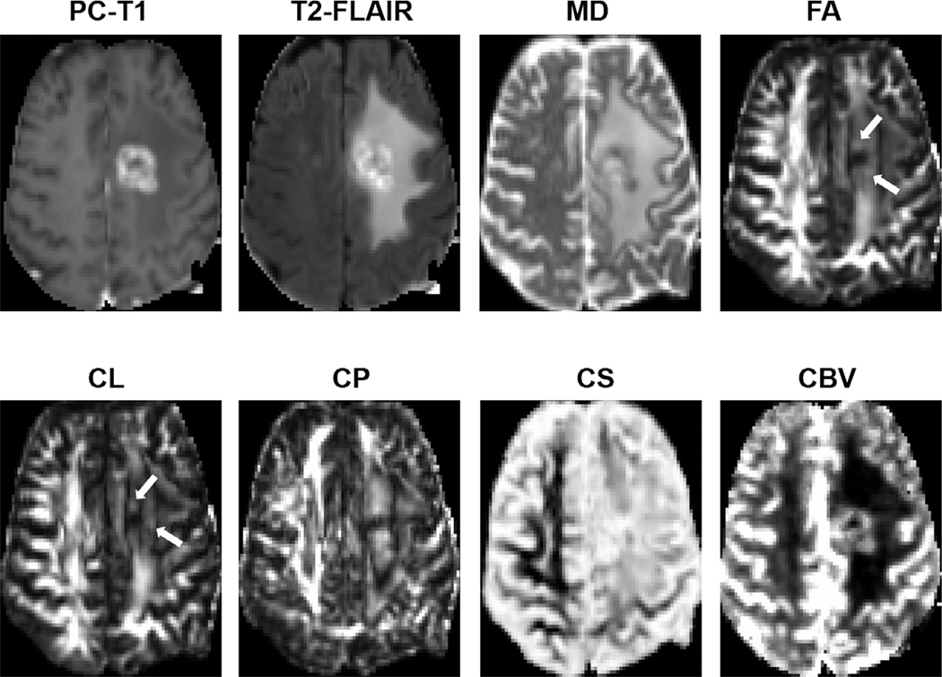
FIGURE 10:
Axial MR images from a 62-year-old patient with TP. Postcontrast T1-weighted image shows an enhancing lesion in the left frontal lobe. Coregistered DTI-derived parametric maps and CBV maps are shown. Increased FA, CL, and CBV are seen corresponding to the areas of enhancement (white arrows).

