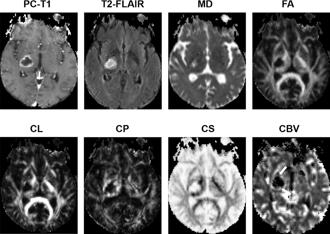
FIGURE 11:
Axial MR images from a 65-year-old patient with PsP. Postcontrast T1-weighted image shows a heterogeneously enhancing lesion in the right thalamic region. Coregistered DTI-derived parametric maps and CBV maps are shown. CBV map shows moderately increased CBV from the lesion (white arrows). Decreased FA, CL, and CP and increased CS are observed from the enhancing part compared with normal white matter. Also, note the presence of lower CBV from contrast-enhancing regions compared to that from the TP patient shown in Fig. 10, suggesting a lower degree of perfusion and neovascularization in PsP compared to TP.

