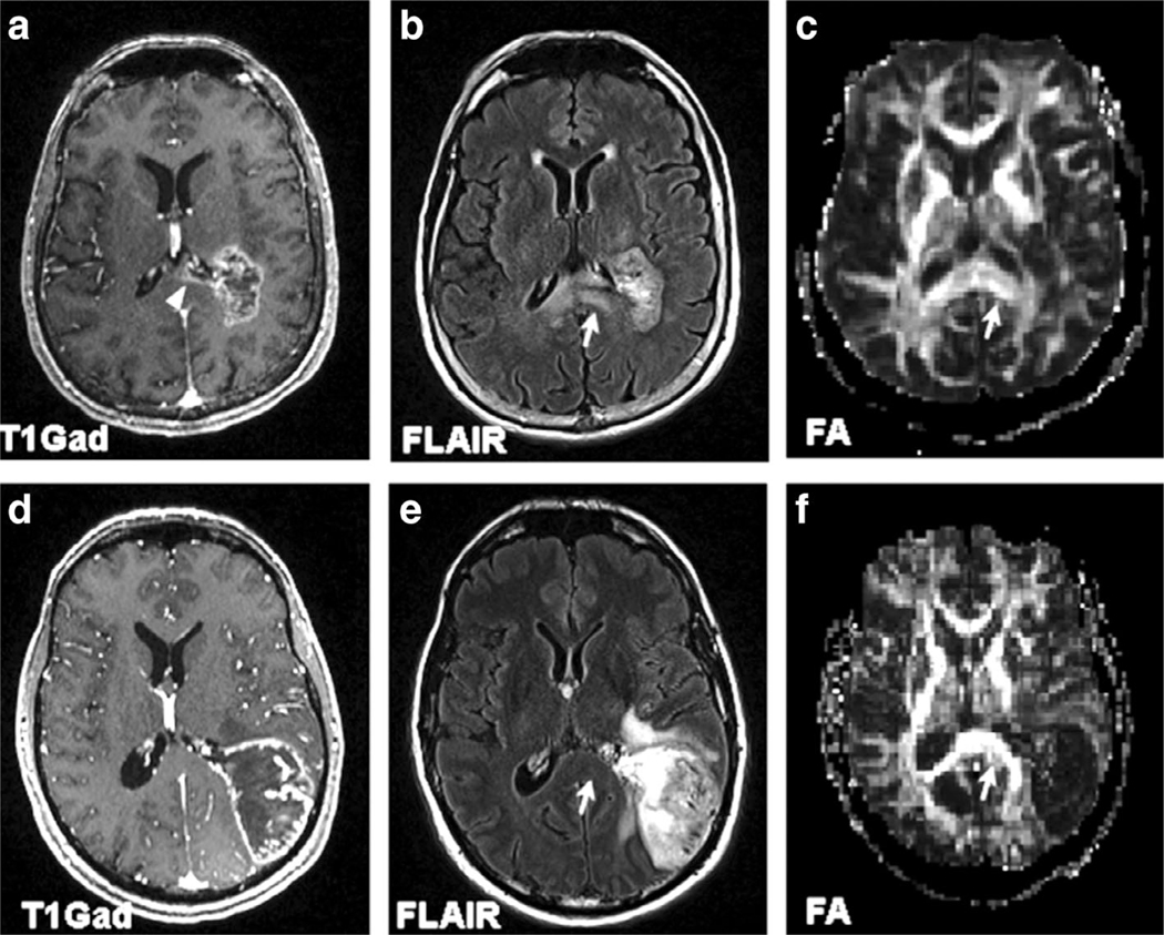
FIGURE 2:
MR images of 70-year-old female glioblastoma patient with corpus callosum invasion (top panel) (a–c) and a 53-year-old male glioblastoma patient without corpus callosum invasion (bottom panel) (d–f). Axial contrast-enhanced T1-weighted images (a,d) demonstrate the extension of enhancing tumor into the splenium of CC in (a) (arrowhead), with normal-appearing CC in (d). Axial FLAIR images (b,e) demonstrate corresponding expansile infiltrative FLAIR signal abnormality in the splenium of corpus callosum (arrow) in (b), whereas the CC seems uninvolved in (e, arrow). Lower FA from the splenium of the corpus callosum is noticed in the FA map (c, arrow) for the patient with shorter survival than the one with longer overall survival (f, arrow). Reprinted with permission from Ref. 39 (Mohan et al., Eur J Radiol 2019 Mar; 112:106–111).

