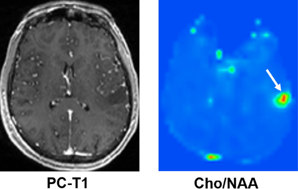
FIGURE 4:
Axial postcontrast T1-weighted image demonstrating a nonenhancing neoplasm in the left parietal lobe; however, the corresponding 3D-EPSI derived metabolite ratio map demonstrates high Cho/NAA, suggesting a higher-grade glioma. On histopathology (not shown), this neoplasm showed areas of increased mitotic activities and pseudopalisading necrosis consistent with GBM (WHO grade IV).

