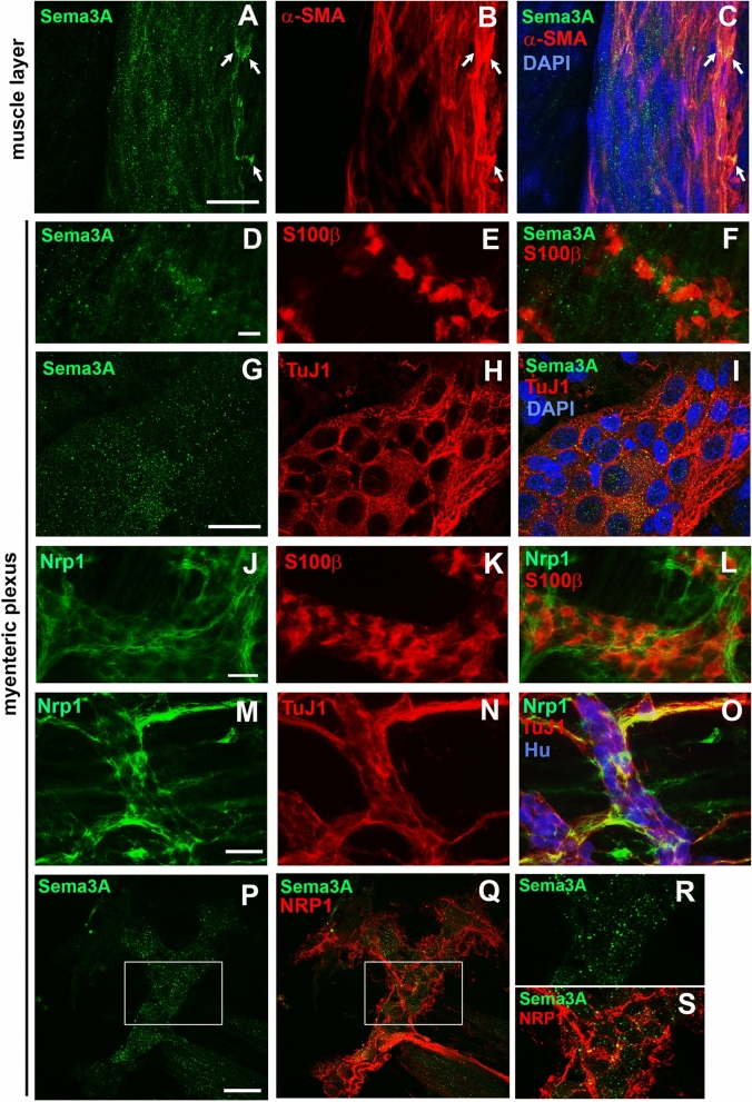
Figure 2.
Sema3A and NRP1 are both associated with ganglia in the myenteric plexus of the distal colon in 7- and 36-day-old rats. (A–C) Double immunolabeling for Sema3A (A,C in green) and α-SMA (marker for muscle cells; B,C in red) in the muscle layer of 7-day-old rat distal colon. Cell nuclei are labeled with DAPI. Sema3A immunostaining appears as numerous small puncta throughout the tissue, closed to muscle cells. In some instances, a higher concentration of Sema3A labeling, more uniformly distributed, is associated with muscle cells (arrows). (D–F) Double immunolabeling for Sema3A (D,F in green) and S100β (marker for enteric glial cells; E,F in red) in the myenteric plexus of 7-day-old rat distal colon. (G–I) Double immunolabeling for Sema3A (G,I in green) and TuJ1 (marker for neuronal soma and axonal processes; H,I in red) in the myenteric plexus of 7-day-old rat distal colon. Cell nuclei are labeled with DAPI. (J–L) Double immunolabeling for NRP1 (J,L in green) and S100β (K,L in red) in the myenteric plexus of 7-day-old rat distal colon. (M–O) Triple immunolabeling for NRP1 (M,O in green), TuJ1 (N,O in red) and Hu (marker for neuronal soma, O in blue) in the myenteric plexus of 7-day-old rat distal colon. (P,Q) Double immunolabeling for Sema3A (P, in green) and NRP1 (Q, in red) in the myenteric plexus of 36-day-old rat distal colon. (R,S) Higher magnification of the boxed regions in (P,Q). Scale bars A–C,G–I 15 µm; D–F,J–Q 50 µm.

