
Fig. 1.
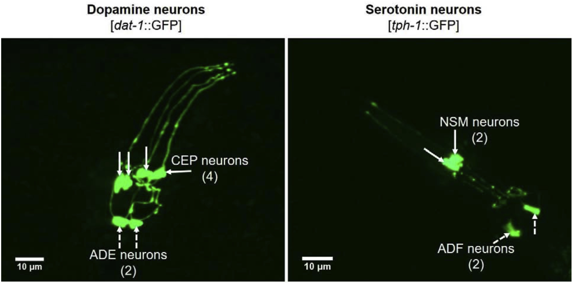
Confocal imaging of DA and SER neurons in the head region of dat-1::GFP and tph-1::GFP C. elegans. Representative images were captured by confocal microscopy (PerkinElmer Spinning disc, Inverted Nikon TE2000-S). CEP – cephalic neurons; ADE – anterior deirid neurons; NSM – neurosecretory motor neurons; ADF – amphid neurons.