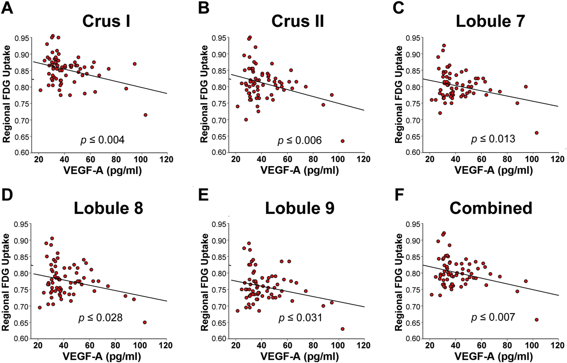
Fig. 2.

Higher plasma VEGF-A levels correspond with lower regional FDG-uptake in blast-mTBI veterans.
Resting state [18F]-fluorodeoxyglucose (FDG) PET imaging in veterans with blast-related mTBI (N = 68) was significantly negatively correlated with levels of plasma VEGF-A in cerebellar VOIs comprising left + right (A) Crus I; (B) Crus II; (C) Lobule 7; (D) Lobule 8; (E) Lobule 9; and (F) combined (Crus I, II, Lobules 7–9), indicating that increasing plasma VEGF-A levels corresponded with reduced metabolism in the cerebellum as measured by FDG-PET.
