Figure 2:

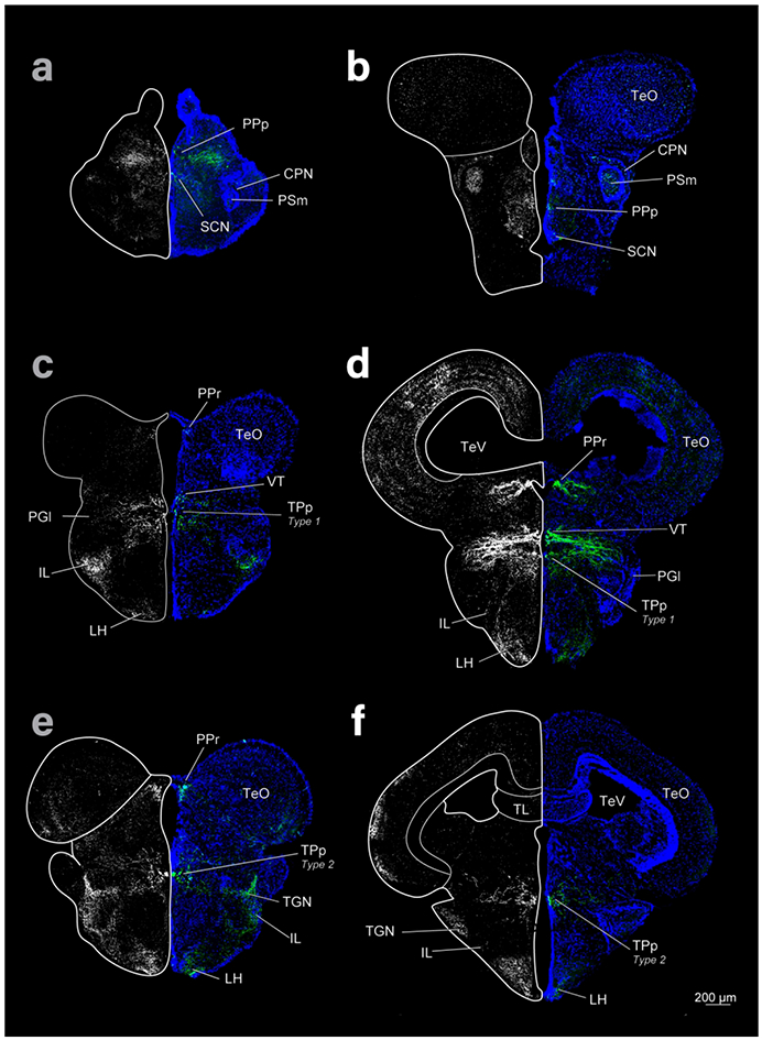
Diencephalon and mesencephalon of cave (left column) and surface (right column) Astyanax showing THir labeling (grayscale images and green) and DAPI (blue) in coronal sections. Top row is rostral, bottom caudal.

Diencephalon and mesencephalon of cave (left column) and surface (right column) Astyanax showing THir labeling (grayscale images and green) and DAPI (blue) in coronal sections. Top row is rostral, bottom caudal.