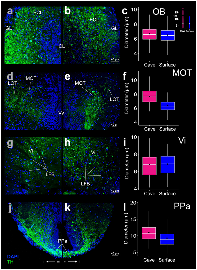
Figure 6:

THir (green) and DAPI (blue) stained coronal sections of cave (left; a, d ,g, j) and surface (right; b, e, h, k) Astyanax. THir labeled somata had significantly larger (p-value < 0.001) diameters in cave Astyanax than in surface Astyanax in the olfactory bulb (OB; a-c), medial olfactory tract (MOT; d-f), and anterior parvocellular preoptic nucleus (PPa; j-k) but not in the intermediate nucleus of the ventral telencephalic area (Vi; g-i). The box plots in the right column (c, f, i, l) show the distributions of THir somata diameters of cave (pink) and surface (blue) Astyanax for that row. Black dots are the mean diameter, middle white line is the median, limits of the colored box indicate quartiles, and vertical white lines extend to the minimum and maximum diameters. The inset in (c) shows the distribution of OB THir somata diameters of cave (pink) and surface (blue) Astyanax. Dotted line is the threshold for separation of two populations of neurons in cavefish.
