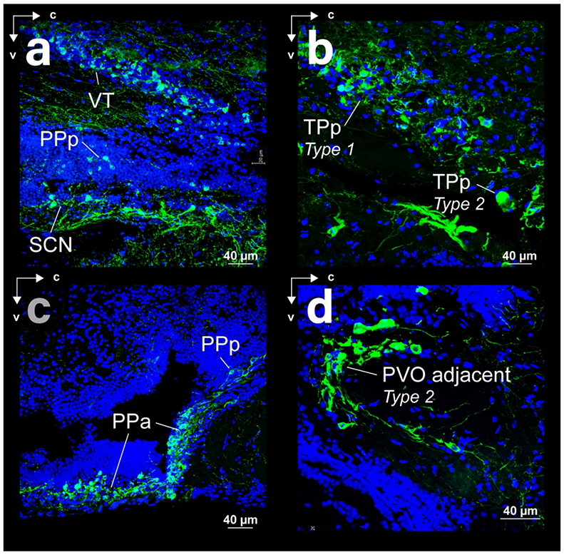
FFigure 7:

Spatial relations of somata and fibers in sagittal sections with THir (green) and DAPI (blue) staining. (a) Ventral thalamus (VT) and preoptic area of surface Astyanax (posterior parvocellular preoptic nucleus - PPp and suprachiasmatic nucleus - SCN). (b) Periventricular nucleus of the posterior tuberculum of surface Astyanax. (c) Anterior (PPa) and posterior (PPp) regions of the parvocellular reoptic nucleus of cave Astyanax. (d) Magnocellular type 2 neurons of the posterior tuberculum, adjacent to the periventricular ventral organ (PVO) of surface Astyanax.
