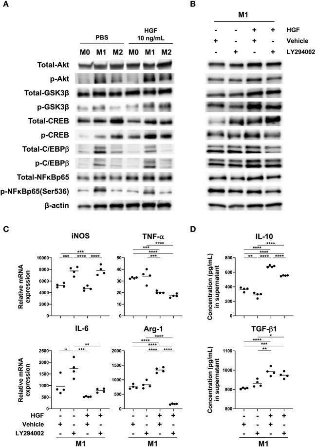
Figure 3.
HGF-MET signaling induces PI3K-mediated Arg-1 expression. (A) Effect of HGF treatment on activity of the PI3K pathway in macrophages. Bone marrow-derived macrophages (BMDMs; M0 macrophages) were induced through the treatment of bone-marrow cells with M-CSF (25 ng/mL) for 7 days. Macrophages were polarized to M1 and M2 phenotypes with treatment for 48 h using IFN-γ and LPS or IL-4, respectively. Western blotting was used to analyze the levels of total and phosphorylated Akt, GSK-3β, CREB, C/EBPβ, and NF-κB. (B) Effects of HGF treatment on downstream PI3K signaling in the presence and absence of PI3K inhibitor. M1 macrophages were treated with a PI3K inhibitor (LY294002) for 1 h after PBS or HGF treatment for 24 h. Western blotting was used to analyze the levels of total and phosphorylated Akt, GSK-3β, CREB, C/EBPβ, and NF-κB. β-actin was used as a loading control. (C) Effect of PI3K inhibitor treatment on the expression of M1 and M2 markers with or without HGF treatment. RT-qPCR was used to compare mRNA expression of iNOS, TNF-α, IL-6, and Arg-1 in M1 macrophages that were treated with or without PI3K inhibitor and HGF for 24 h. The Ct values were normalized to those of β-actin and are expressed relative to mean levels in M0 macrophages not treated with HGF, which is arbitrarily defined as 1. (D) ELISA was used to measure the secretion of IL-10 and TGF-β1. Data are represented as the mean and a scatterplot showing individual data points (n = 4). One-way ANOVA, *P < 0.05, **P < 0.01, ***P < 0.001, and ****P < 0.0001.

