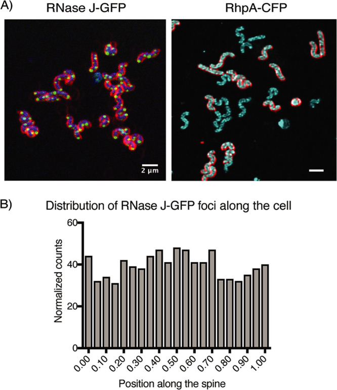
FIG 2.

RNase J and RhpA form foci in H. pylori cells that do not have a polar localization. (A) Representative composite confocal microscopy images of live H. pylori cells expressing RNase J-GFP (green) or RhpA-CFP (cyan). In the RNase J-GFP image, blue indicates DNA (Hoechst 33342); in both images, red indicates membranes (FM4-64). Experiments were performed at least 3 times. (B) Histogram showing the number of foci of RNase J-GFP that are located in each position along the spine of the H. pylori cells.
