FIG 7.
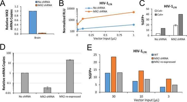
Depletion of MX2 partially relieves blocks to HIV-1 infectivity. P. alecto kidney, fetus, and brain cells were transduced with lentiviral vectors encoding mCherry and an shRNA cassette specific for bat MX2. (A) Stably transduced cells were analyzed for MX2 transcript expression by qPCR normalized to GAPDH. (B) Wild-type and MX2 knockdown brain cells were challenged with a luciferase-encoding HIV-1LTR vector, and 3 days postinfection, cells were counted, lysed, and analyzed for luminescence. RLU data were normalized to the number of healthy cells and are shown as average RLU reading ± standard deviation. (C) Wild-type and MX2 knockdown brain cells were challenged with an eGFP-encoding HIV-1LTR vector in the presence of DMSO (CsA-) or 5 μM CsA (CsA+). Cells were cultured for 48 h before GFP was quantified by flow cytometry. (D) MX2 knockdown brain cells were transduced with lentiviral vector encoding a P. alecto MX2 cDNA and a puromycin selection cassette (“back-complement” cells). Puro stable cells were isolated, and MX2 transcripts were quantified as described above. (E) Wild-type, MX2 knockdown, and recomplemented cells were challenged with eGFP-encoding HIV-1LTR and analyzed as described for panel C. The data shown are representative of results from a total of three experiments performed.

