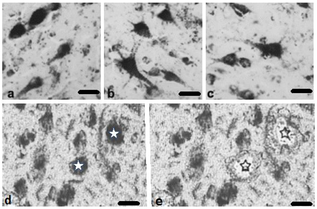
Fig. 1.

Photomicrographs showing PreC CatD-positive layer III neurons in NCI (a), MCI (b), and AD (c). CatD immunolabeled neurons pre (d, white stars) and post (e, black outlines stars) microdissection from a non-cognitively impaired 84-year old female. Distorted appearance of the images (d and e) is due to the fact that the pictures were taken from uncover slipped sections with the aid of a Zeiss PALM III LCM.. Scale bars = 25 μm
