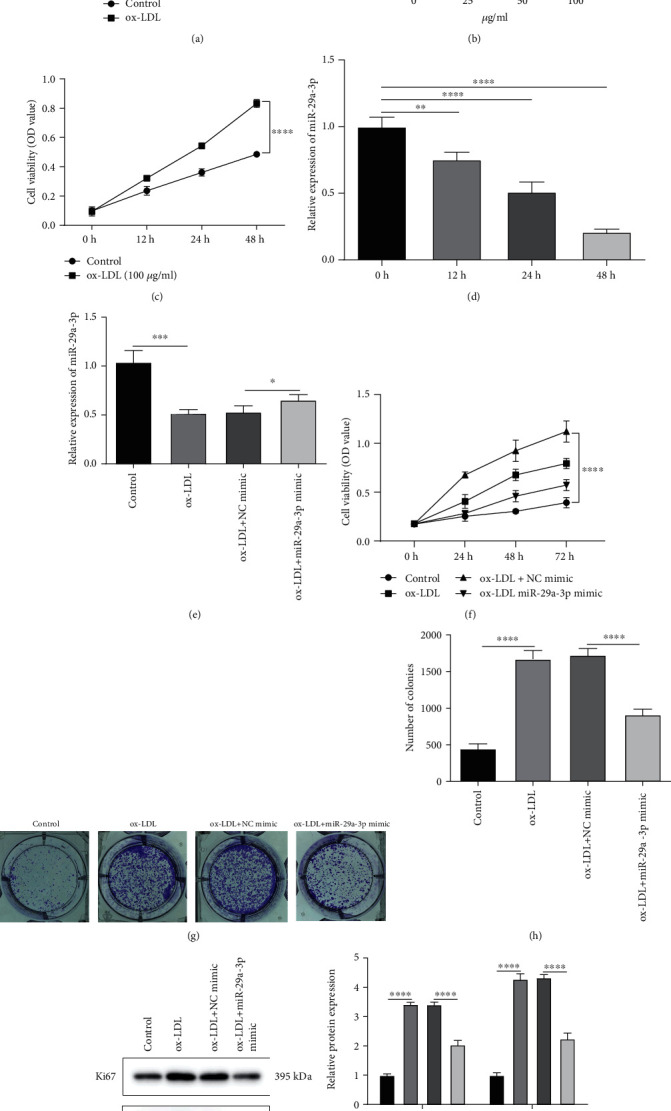
Figure 2.

Overexpression miR-29a-3p suppresses proliferation of ox-LDL-induced vascular smooth muscle cells (VSMCs) in AS. (a) CCK-8 assay was used to detect the cell viability of VSMCs under different concentrations of ox-LDL. (b) miR-29a-3p expression was determined in VSMCs induced by different concentrations of ox-LDL by RT-qPCR. (c) The cell viability of VSMCs induced by 100 μg/ml ox-LDL was detected using CCK-8 assay in different time periods. (d) RT-qPCR was presented to examine the expression of miR-29a-3p in VSMCs induced by100 μg/ml ox-LDL in different time periods. (e) miR-29a-3p expression was tested in ox-LDL-induced VSMCs with or without miR-29a-3p mimic transfection by RT-qPCR. (f) CCK-8 assay was presented in ox-LDL-induced VSMCs with or without miR-29a-3p mimic transfection. (g, h) Colony formation assay was used to examine the proliferative ability of ox-LDL-induced VSMCs after transfection with miR-29a-3p mimic or not. (i, j) The expression of Ki67 and PCNA proteins was examined in ox-LDL-induced VSMCs with or without miR-29a-3p mimic transfection. ∗p < 0.05; ∗∗p < 0.01; ∗∗∗p < 0.001; ∗∗∗∗p < 0.0001.
