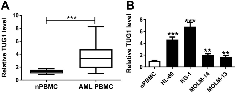
Figure 1.
Expression of TUG1 in acute myeloid leukemia (AML) specimens and cells. (A and B) TUG1 expression levels were detected by RT-qPCR analysis in (A) the peripheral blood mononuclear cells (PBMC) derived from AML patients (AML nPBMC; n=23) compared with the healthy normal PBMC (nPBMC; n=12), and (B) human AML cell lines (HL-60, KG-1, MOLM-14, and MOLM-13) versus nPBMC. The error bars represent the mean±standard error of mean (SEM) of three independent experiments. **P<0.01, and ***P<0.001.

