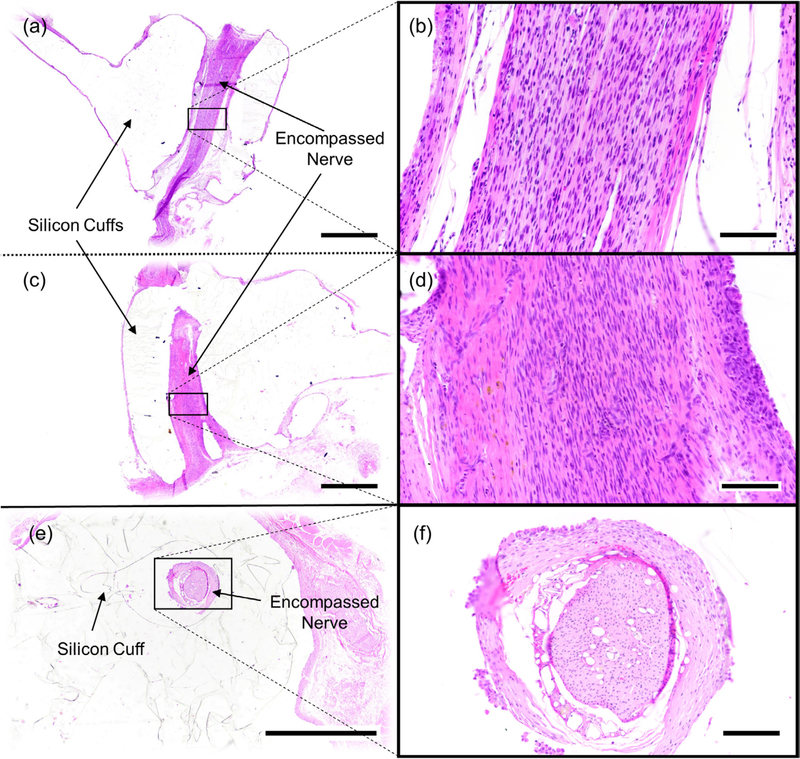
Figure 6.
Representative photomicrographs of cuffed vagus nerves stained with hematoxylin and eosin (HE). Images ((a)–(d)) are from the same nerve (eRx143). Images ((e) and (f)) are of a separate nerve (eRx132). (a) Longitudinally sectioned nerve encompassed by a stimulation cuff, (b) higher magnification of (a). (c) Longitudinally sectioned nerve encompassed by a caudally positioned recording cuff, (d) higher magnification of (c). (e) Example of a nerve cuffed with an inert cuff with no metal electrodes sectioned in the transverse plane, (f) higher magnification of (e). Scale bars = 1000 microns ((a), (c) and (e)) or 100 microns ((b), (d) and (f)).

