Figure 5.

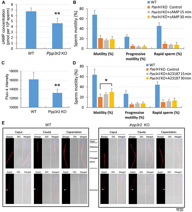
Loss of PPP3R2 function reduces cAMP and calcium levels in sperm and compromises membrane diffusion during sperm maturation. (A) cAMP concentration declines in Ppp3r2 KO sperm. Error bars represent SD (n = 8). **P < 0.01. (B) db-cAMP supplementation fails to reverse sperm motility decrement in Ppp3r2 KO sperm. Error bars represent SD (n = 7). (C) Calcium level, determined by Fluo-4 staining and flow cytometry, decreases in Ppp3r2 KO sperm. Error bars represent SD (n = 8). **P < 0.01. (D) A23187 treatment fails to restore Ppp3r2 KO sperm motility to a normal level of WT sperm even though transiently increased for 30 min. Error bars represent SD (n = 7). *P < 0.05. (E) Immunofluorescent staining of Basigin and Sept4 in sperm during epididymal transit and capacitation: in WT mice, Basigin is localized in the principal piece of caput epididymis, and after epididymal transit, its localization is restricted to the midpiece of cauda epididymis sperm. Ultimately, it is localized in the whole tail after capacitation, whereas in the Ppp3r2 KO mice, Basigin localization pervades throughout the whole tail of cauda epididymis sperm. Sept4 expression is limited to the annulus (arrows) in WT sperm but is dispersed in Ppp3r2 KO sperm. These results suggest that loss of diffusion barrier integrity occurs specifically in the cauda epididymis. Scale bar, 10 μm.
