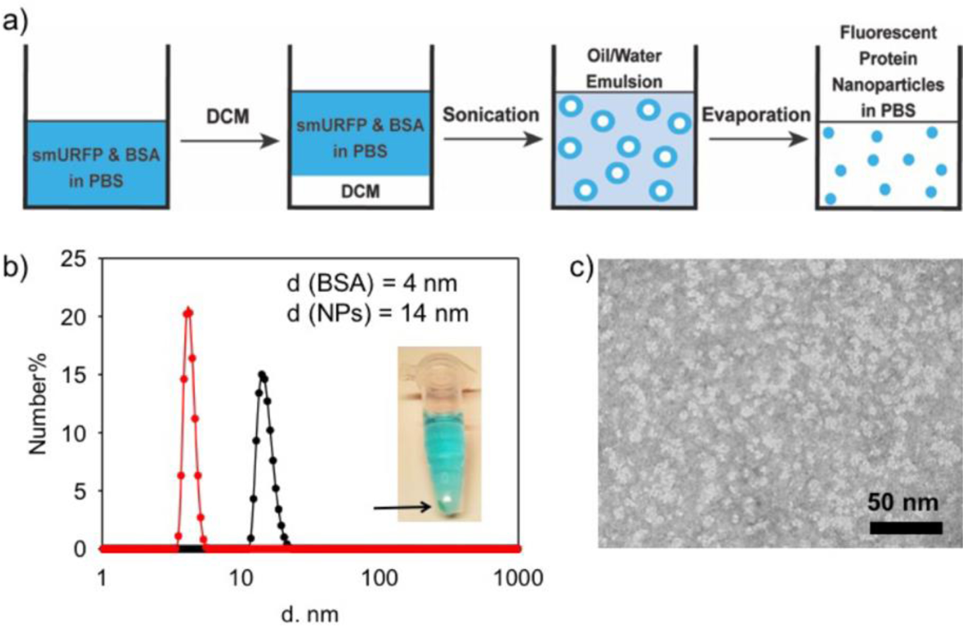
Figure 1.

Synthesis and size characterization of fluorescent protein nanoparticles. a) Schematic of fluorescent protein nanoparticle preparation by the emulsion method. b) The DLS characterization of BSA and the fluorescent protein nanoparticles (NPs). Diameter (d) is shown on the X-axis with a log scale. Inset: Centrifuged Eppendorf tube showing precipitated protein in the pellet and fluorescent protein nanoparticles in solution. c) TEM image of fluorescent protein nanoparticles negatively stained with 0.5% uranyl acetate. Fluorescent protein nanoparticles are shown as white in the image.
