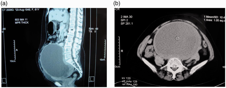
Figure 1.
Abdominal computed tomography revealed a capsulated cystic mass (18 × 14.5 × 14 cm) that encompassed the hypogastric region and pelvis. (a) Median sagittal section. (b) Coronal section. The mass exhibited a close relationship to the bladder, ureter, and rectum. No obvious bony destruction, defect, or resorption was seen.

