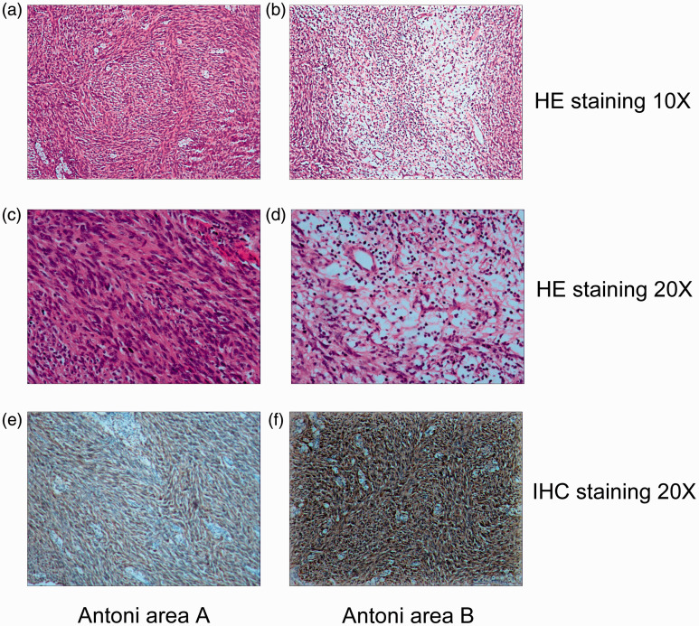
Figure 3.
Histopathology of retroperitoneal neurilemmoma. The mass consisted of a proliferation of fusiform cells that formed a palisade or turbinate pattern ((a) ×10, (c) ×20) and of myxoid and degenerated tissue with cells and gelatinous substance ((b) ×10, (d) ×20). (e, f) S-100-positive cells (×20).

