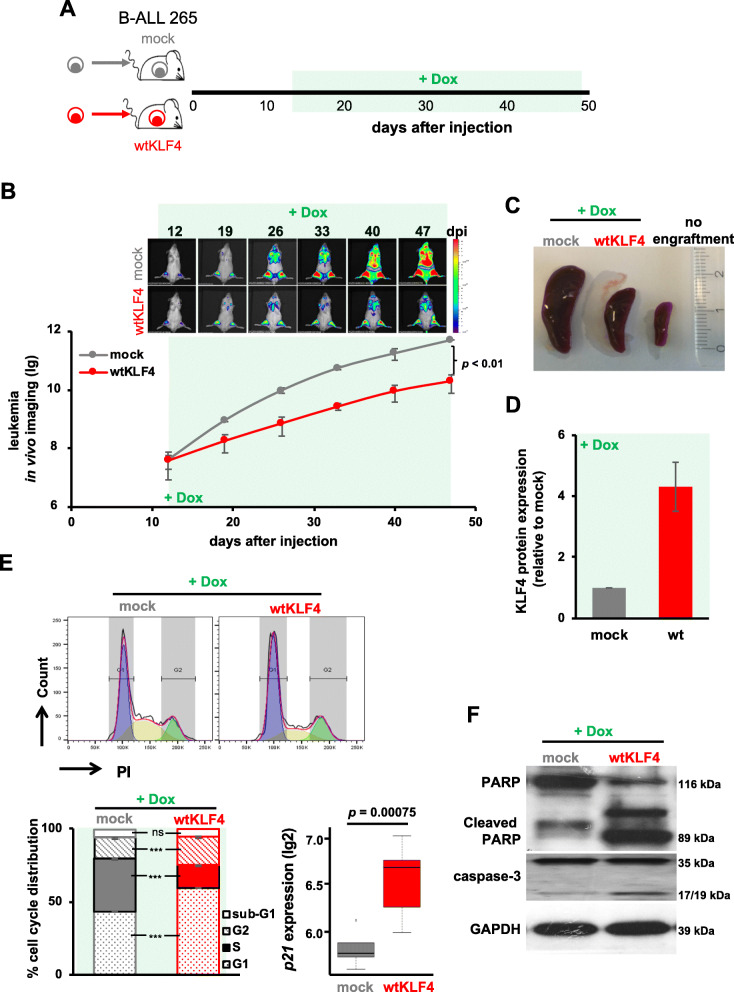
Fig. 3.
Re-expressing KLF4 inhibits B-ALL PDX growth through cell cycle arrest and apoptosis in vivo. a Experimental design: Sixty thousand triple-transgenic, mock or wtKLF4 PDX ALL-265 cells were injected into the tail vein of 6 NSG mice each. After homing was completed and the tumors were established, Dox (1 mg/ml) was added to the drinking water from day 12 onwards to induce KLF4 expression. On day 47 after cell injection, the mice were sacrificed, spleens harvested and the Dox-induced, mCherry-positive mock- or wtKLF4-expressing cell population was enriched by flow cytometry for further analysis. b In vivo bioluminescence imaging is shown as representative images (upper panel), and after the quantification of all 6 mice, the results are depicted as the mean ± SEM. p < 0.01 according to a two-tailed unpaired t test. c Representative spleens of mice, with a healthy mouse without leukemic engraftment for comparison. d The KLF4 protein level of mCherry-positive splenic cells was analyzed by capillary immunoassays; β-Actin served as the loading control. The mean ± SD of KLF4 expression normalized to that of β-Actin for 3 mice is plotted as the fold change relative to the mock control. e Cell cycle analysis: 106 mCherry positive cells were stained with propidium iodide (PI) and cell cycle distribution was measured by flow cytometry. Upper panel: representative histograms of mock- (n = 3) or wtKLF4-expressing (n = 3) cells; lower panel: quantification of the mean ± SD is shown. *** p < 0.005 by two-tailed unpaired t test; ns = not significant. Lower right: Levels of p21 mRNA as determined by scrb sequencing. f PARP- and caspase-3 cleavage in mock- (n = 3) or wtKLF4-transduced (n = 3) PDX ALL-265 cells as determined by Western blotting; GAPDH was used as a loading control

