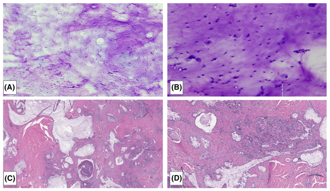
FIGURE 2.

This FNA of a cystic parotid mass demonstrated abundant mucin (A) and macrophages (B) without an evaluable epithelial component, categorized as atypical per MSRSGC. Follow-up revealed a low-grade mucoepidermoid carcinoma (C,D) [Color figure can be viewed at wileyonlinelibrary.com]
