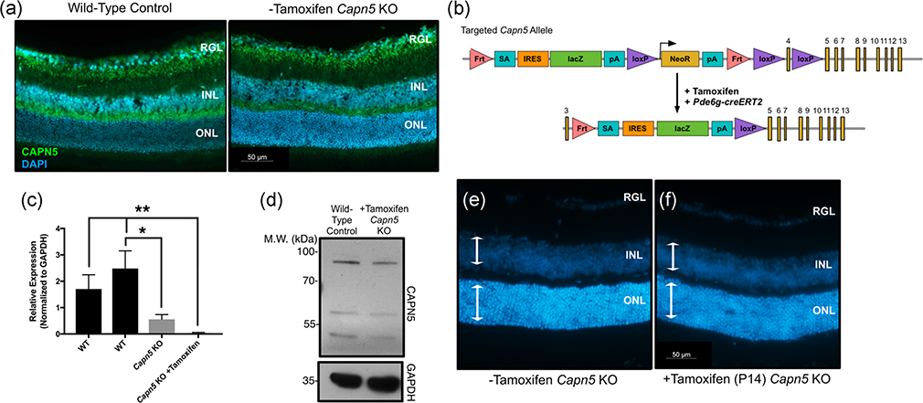
FIGURE 2.
Knockout of Capn5 in the mouse photoreceptor cells leads to no detectable adverse clinical outcomes. (a) 2.5-month-old mouse eyes from wild-type (WT) and Capn5 KO + tamoxifen mice were cross-sectioned from posterior–anterior axis including the optic nerve and stained for CAPN5 protein (green). DAPI (blue) highlights the nuclei. DAPI, 4′,6-diamidino-2-phenylindole; INL, inner nuclear layer; ONL, outer nuclear layer; RGL, retinal ganglion cells. (b) The targeting construct for our Pde6g-creERT2 mouse line (Koch et al., 2015, 2017). One allele of phosphodiesterase 6γ (Pde6g) contains a cassette with creERT2 before exon 4, which is activated by delivery of tamoxifen. 3′-HA, 3′ homology arm; 5′-HA, 5′ homology arm; EM7, bacteriophage T7 promoter; bGHpA, bovine growth hormone polyadenylation signal; NeoR, neomycin resistance; pA, polyadenylation signal; PGK, phosphoglycerate kinase promoter; TK, thymidine kinase. (c) The targeted tm1a allele in Capn5 KO mice after being crossed with the Pde6g-creERT2 mice and after delivery of tamoxifen ensures a loss of the critical Capn5 exon 4. (d) Retinas were dissected at postnatal Day 42 (P42) and genomic DNA was extracted. Quantitative polymerase chain reaction (qPCR) was performed for Capn5 expression in the Capn5 KO mice and Capn5 KO +tamoxifen mice compared to two WT control samples (Far left, 129/SvEV agouti; second to the left, C57BL/6J). Samples were run in triplicate, normalized to Gapdh, and error bars represent the standard deviation between triplicates. The Capn5 KO retina was significantly decreased compared to the C57BL/6J WT sample (p = .0049). The Capn5 KO +tamoxifen retina was significantly decreased compared to both WT samples (p = .0104 and p = .0014, respectively). (e) Western blot analysis of CAPN5 using 2-month-old retinal lysates from WT (left lane) and Capn5 KO +tamoxifen (right lane) mice. GAPDH was used as a loading control. P42 mouse eyes from (f). Capn5 KO mice and G. Capn5 KO + tamoxifen mice were cross-sectioned from posterior–anterior axis including the optic nerve, stained with DAPI to highlight the nuclei, and retinal sections were magnified to examine thickness of retinal cell layers. White arrows, thickness of the INL/ONL, same size across both images. N ≥ 4 mice for all mouse experimental groups. GAPDH, glyceraldehyde 3-phosphate dehydrogenase; INL, inner nuclear layer; KO, knockout; ONL, outer nuclear layer

