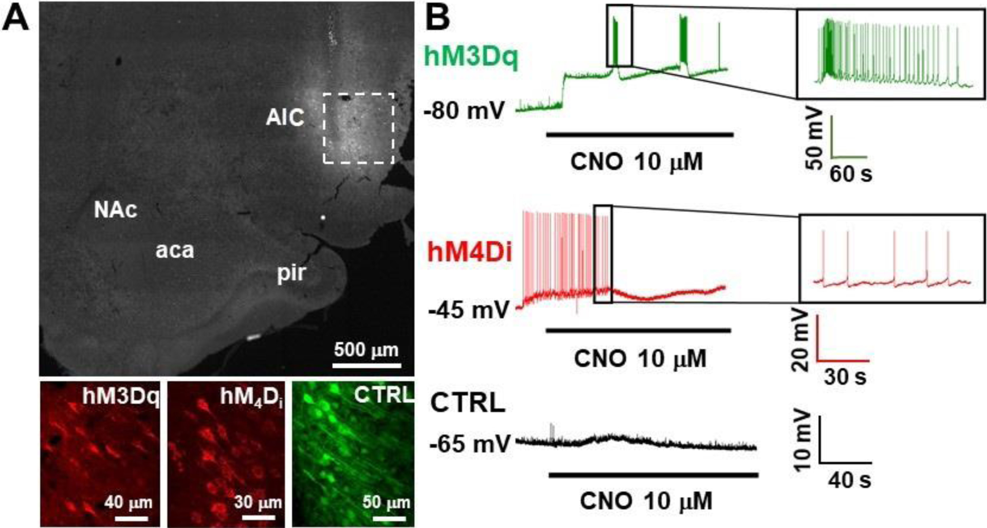
Figure 2.

Validation of DREADD functionality using whole cell patch clamp electrophysiology. A) Representative low magnification images of DREADD expression within the AIC (dashed box), and high magnification images in the AIC of each of the viral vectors utilized (bottom). B) Representative electrophysiological traces for hM3Dq (green; top), hM4Di (red; middle) and control (black; bottom) are shown. Black line below traces represents CNO bath application.
