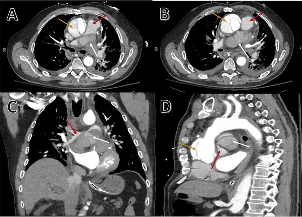
FIGURE 2.

CT angiography of thoracic aorta showing acute Type A aortic dissection. Axial view (A and B), coronal view (C), and sagittal view (D). White arrows show mediastinal hematoma that compresses and obliterates 90% of the bifurcation of pulmonary artery (A–D), red arrows show location of pulmonary artery in relation to mediastinal hematoma, and yellow arrows show location of false lumen (A, B, and D). Mediastinal hematoma extrinsically compressing the pulmonary bifurcation (A–C) giving it a “saddle pulmonary embolism”‐like appearance on CT. Dissection involving the aortic root (D) with defect in posterior wall of proximal ascending aorta forming mediastinal hematoma
