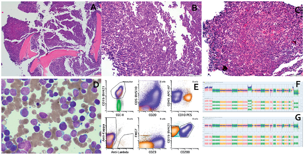
Figure 1.

(a) Low power view showing hypercellular marrow. (b) High power view showing “blastoid” morphology in high grade B-cell lymphoma (HGBCL). (c) High power view showing CLL/SLL. (d) Aspirate smear showing dimorphic population of small lymphocytes with mature chromatin and large lymphoid cells with dispersed chromatin and prominent nucleoli. (e) Flow cytometric plots showing immunophenotype of HGBCL (orange) and CLL/SLL (blue). (f) SNP array findings in HGBCL showing homozygous deletion at 13q14.2 (including RB1 and SETDB2) and CN-LOH of 17p terminal to 17p11.2 (including TP53). (g) SNP array findings in CLL/SLL showing a 13q14.2 hemizygous deletion for RB1 and homozygous deletion for SETDB2 and loss of 17p13.1 to 17p12 (including TP53).
