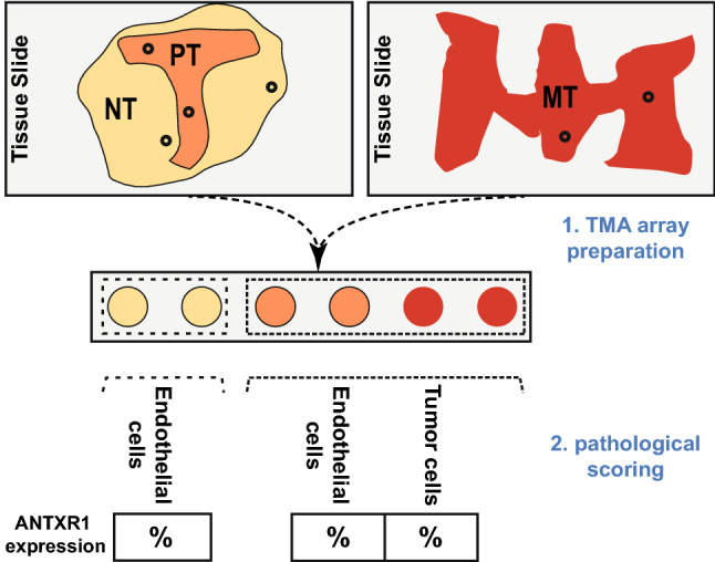
Fig. 1.

Steps from tissue microarray (TMA) preparation to data acquisition. Paraffin blocks of patients with gastric adenocarcinoma were cored according to earlier inspection of their tissue slides with H and E staining. Two cores were collected from each anatomical region, i.e., the non-tumor region (NT), the primary tumor (PT) and the metastatic tumor (MT). Subsequently, after ANTXR1 immunostaining, the proportion of positively stained endothelial and tumor cells among all endothelial and tumor cells in each core was reported as a percentage
