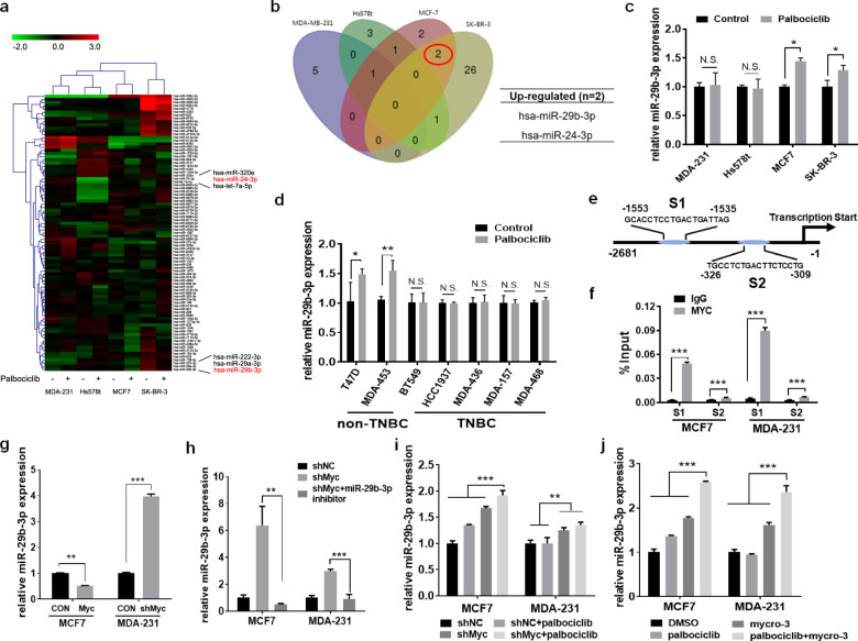
Fig. 3. Palbociclib-induced miR-29b-3p is negative regulated by c-myc.
a A miRNA microarray was performed to detect differentially expressed miRNAs of MDA-MB-231, Hs578t, MCF-7, and SK-BR-3 cells treated with or without 4 μM palbociclib for 48 h. The green in the legend represents downregulation, and the red represents upregulation (>1.5-fold change in expression, P < 0.05). b Venn diagrams for the number of differentially expressed miRNAs among four breast cancer cell lines after the treatment of palbociclib for 48 h. c Validation by quantitative real-time PCR (qRT-PCR) of miR-29b-3p in the four breast cancer cells treated with or without palbociclib. d miR-29b-3p in the other seven breast cancer cells treated with or without palbociclib were analyzed by qRT-PCR. U6 was used as an endogenous control for miRNA analysis. e Schematic model of c-myc binding sites in S1 promoter and S2 promoter of miR-29b-3p gene by bioinformatic analysis. The sites and sequences of three binding sites were indicated in model scheme. f Binding of c-myc in MCF-7 and MDA-MB-231 cells to the miR-29b-3p promoter region was analyzed by CHIP-qPCR. g qRT-PCR analysis of miR-29b-3p expression in MCF-7 and MDA-MB-231 cells after overexpression or knockdown of c-myc. Expression of miR-29b-3p in MCF-7 and MDA-MB-231 cells transfected with NC shRNA or c-myc shRNA treated with h miR-29b-39 inhibitor or i palbociclib was analyzed by qRT-PCR. j Expression of miR-29b-3p in MCF-7 and MDA-MB-231 cells following treatment with the indicated drugs was measured by qRT-PCR. Error bars indicate mean ± standard deviation.

