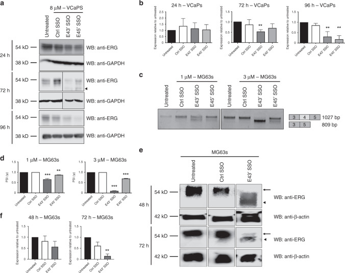
Fig. 2. Splice-switching caused by ERG exon 4 SSOs reduces ERG protein levels in two cancer cell lines.
a Representative ERG and GAPDH (loading control) western blots of lysates from VCaP cells treated with 8 µM SSOs for 24, 72 and 96 h. The arrowhead indicates a putative truncated ERG isoform (full length is 54kD). b Quantifications of ERG western blots from VCaP cells treated with SSOs for 24–96 h (n = 4 for 24 h and 72 h, n = 3 for 96 h, except for ctrl SSO at 96 h, n = 2). c Representative RT-PCR panels for MG63 cells treated with 1 or 3 µM for 24 h. d Quantification of exon 4 skipping in MG63 cells treated with SSOs at 1 or 3 µM for 24 h (n = 3). e Representative ERG and β-actin (loading control) western blots of lysates from MG63 cells treated with SSOs for 48 and 72 h. The arrow indicates full-length ERG isoform (54 kD) and arrowhead indicates truncated ERG isoform. f Quantifications of ERG western blots from MG63 cells treated with 5 µM SSOs for 48 and 72 h (n = 8 for untreated and ctrl SSO, for E43′ n = 4 at 48 h; n = 6 at 72 h). ERG protein expression levels were normalised to GAPDH. *** = p < 0.001, ** = p < 0.01, * = p < 0.05. Ctrl SSO control SSO.

