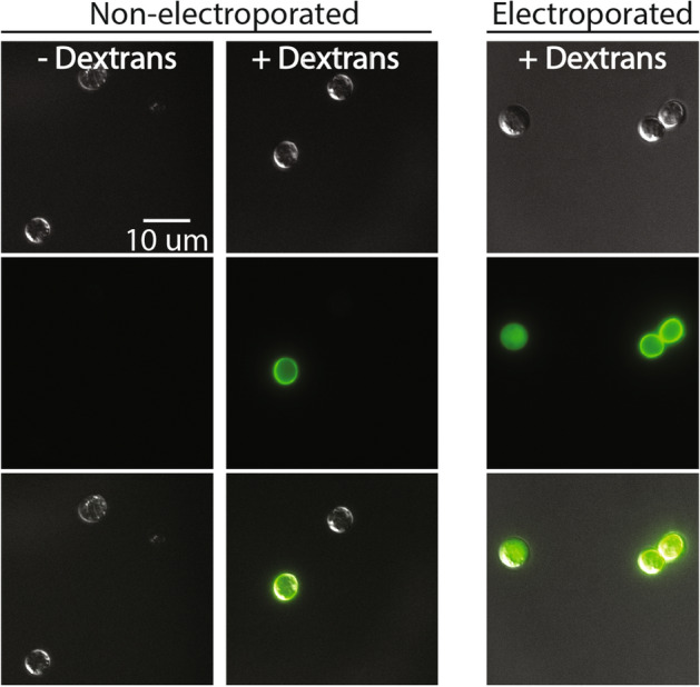
Figure 5.

Although dextran staining of cell wall/coat occurs without electroporation, stained and electroporated cells can be distinguished via microscopy. Representative images of each treatment were taken from a single replicate performed on subsets of spores from the same population. Fluorescence intensity was normalized across all images. Non-electroporated cells incubated with dextrans (non-electroporated, + dextrans) show weak staining in a pattern that suggests binding to the cell wall. This same pattern can be observed in electroporated samples (electroporated, + dextrans) where stained cells (right) co-occur with positive cells (left) in which dextrans fluorescence is limited to the inside of the cell.
