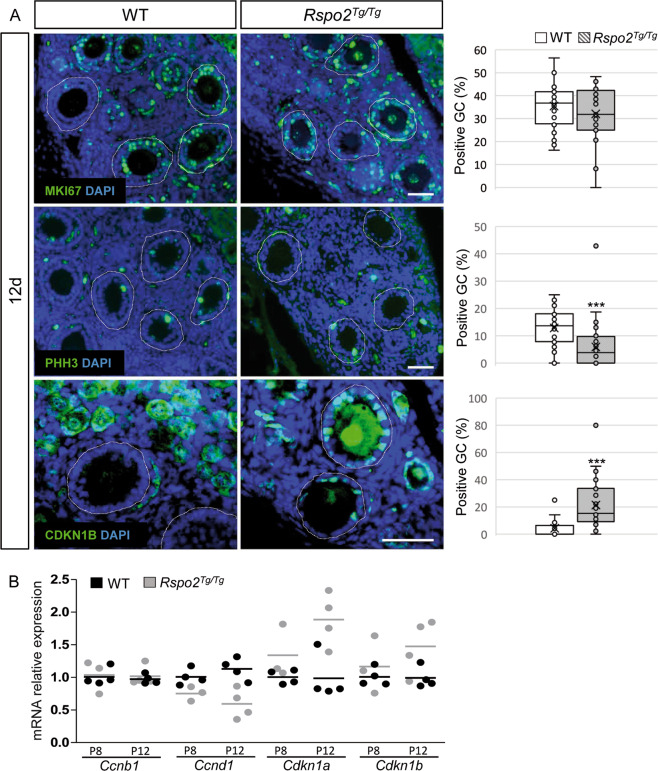
Fig. 3. Granulosa cell cycle progression is impaired in the absence of RSPO2.
a Immunodetection and quantitative analyses of MKI67, PHH3, and CDKN1B (p27) to assess the proliferation status of granulosa cells at 12d in WT and Rspo2Tg/Tg transplanted ovaries. Left panel: follicular cells are engaged in cell cycle as indicated by MKI67 immunostaining but exhibit an interphase delay illustrated by a decrease of PHH3-positive and an increase of CDKN1B-positive granulosa cells in mutant ovaries. Follicles are outlined with a white dotted line. Right panel: quantification of the % of positive granulosa cells for MKI67, PHH3, and CDKN1B. n = 1070, 1107, and 280 WT and n = 889, 678, and 583 Rspo2Tg/Tg granulosa cells (identified with DAPI) from 31, 30, and 10 and 28, 42, and 23 follicles, respectively, from at least two individual transplanted ovaries of each genotype. Data are presented in a box and whisker plot representation to illustrate positive granulosa cell dispersion according to the follicle considered. Student’s t test, unpaired two sided (*p < 0.05; **p < 0.01; ***p < 0.001). Scale bar, 50 µm. b QRT-PCR analysis of Ccnb1 (CyclinB1), CcnD1 (CyclinD1), Cdkn1a (p21), and Cdkn1b (p27). Both inhibitors of CDK are upregulated, while Ccnd1 expression is decreased, confirming a reduction of the cell cycle progression in Rspo2Tg/Tg granulosa cells. Data are presented as individual data points. n = 3 (8d) and 4 (12d) individual ovaries per genotype. Mean values are indicated as black (WT) and gray (Rspo2Tg/Tg) bars.

