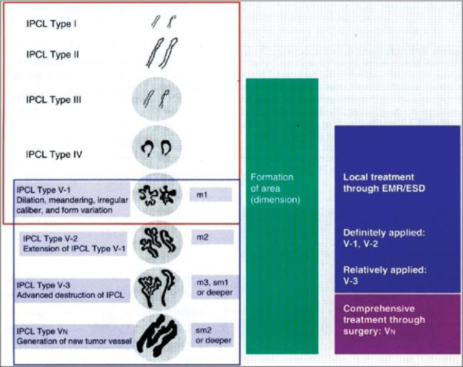
Figure 2:
Original intrapapillary capillary loop (IPCL) pattern classification.
The IPCL pattern classification includes two sets of diagnostic criteria. IPCL pattern classification from IPCL type I to type V-1 is used for the tissue characterization of flat lesions (red outline). IPCL pattern classification from IPCL type V-1 to type VN reflects cancer infiltration depth (blue outline). IPCL type III corresponds to borderline lesions which potentially include esophagitis or low-grade intraepithelial neoplasia. IPCL type III should be considered for endoscopic follow up. In IPCL type IV, high-grade intraepithelial neoplasia appears, and then further treatment with endoscopic mucosal resection (EMR) / endoscopic submucosal dissection is recommended. EMR/esophageal squamous dysplasia for IPCL types V-1 and V-2 should be also considered as they are definite M1 or M2 lesion with no risk of lymph node metastasis. IPCL type V3 corresponds to an M3 lesion and diagnostic EMR/esophageal squamous dysplasia should be applied as a “complete biopsy” to decide on a final treatment strategy. IPCL type VN corresponds to a “new tumor vessel” often associated with sm2 invasion with significantly increased risk of lymph node metastasis. Surgical treatment should be recommended.
(Reprinted with permission from Inoue H, et al. Ann Gastroenterol. 2015 Jan-Mar; 28(1): 41–48.)

