Figure 4:
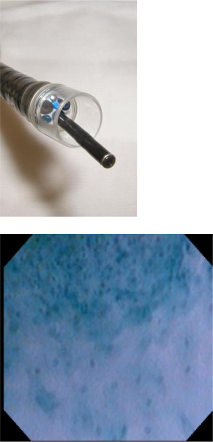
Microendoscopic imaging of squamous dysplasia.
a. | Endocytoscope probe which can be passed through the instrument channel of an endoscope.
b. | The border between esophageal squamous cancer (upper) and normal squamous epithelium (lower). The density and nucleus:cytoplasm ratio is much higher in cancer compared to normal epithelium.
(Reprinted with permission from Kumagai Y, et al. Endoscopy 2004;36:590–594.)

