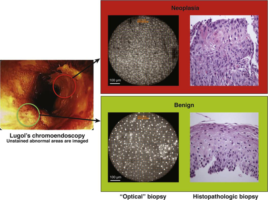
Figure 5:
High-resolution microendoscope92
Lugol’s unstained lesions (left) are imaged with HRME (“optical” biopsy) and compared with corresponding tissue biopsy (histopathologic biopsy) (original magnification, 100x). Only 1 of the 2 Lugol’s abnormal areas was neoplastic (upper panel) as determined by the imaging software, based on mean nuclear area, nuclear-to cytoplasmic ratio, nearest inter-nuclear distance, nuclear eccentricity, nuclear solidity, and the major axis of the ellipse best approximating each nucleus.
(Reprinted with permission from Protano et al. Gastroenterology 2015; 149: 321–329.)

