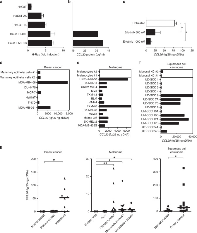
Fig. 1. Oncogenic Ras induces CCL20 protein synthesis.
a Relative Ras activity in the immortalised keratinocyte cell line HaCaT and in H-RasV12-transfected HaCaT clones was assayed by the EZ-Detect Ras activation kit. b CCL20 protein production- conditioned medium from HaCaT cells and H-RasV12-transfected HaCaT clones, as detected by a CCL20-specific ELISA. c Activated primary keratinocytes were treated with the selective irreversible inhibitor of EGFR erlotinib, and expression of CCL20 was analysed by qPCR. Tumour cells derived from breast cancer, malignant melanoma and head and neck squamous cell carcinoma (HNSCC) overexpress CCL20. d–f Quantitative real-time PCR analysis of CCL20 in cultured normal primary mammary epithelial cells (n = 2) and breast cancer cell lines (n = 7) (d), cultured normal primary melanocytes (n = 2) and melanoma cell lines (n = 13) (e), cultured primary mucosal keratinocytes (KC, n = 2), cell lines derived from primary tumours (n = 10) or metastases (n = 4) of HNSCC (f). g CCL20 expression of tumour tissues derived from breast cancer (primary breast cancer, n = 12, fc = 1.096; breast cancer metastasis, n = 10, fc = 6.048), malignant melanoma (primary melanoma, n = 28, fc = 4.580; subcutaneous metastasis, n = 11, fc = 6.023; distant metastasis, n = 4, fc = 6.012) and HNSCC (primary tumour, n = 14, fc = 3.057) compared with normal tissue (normal breast, n = 3, cultured primary melanocytes, n = 3, benign nevi, n = 5 and normal oral mucosa, n = 8) by qPCR. Values are either expressed as femtograms of target gene per 25 ng of cDNA or protein concentration in picograms per ml of supernatant and represent the mean ± SD of three independent experiments (*P ≤ 0.05; **P ≤ 0.01; Mann–Whitney U test).

