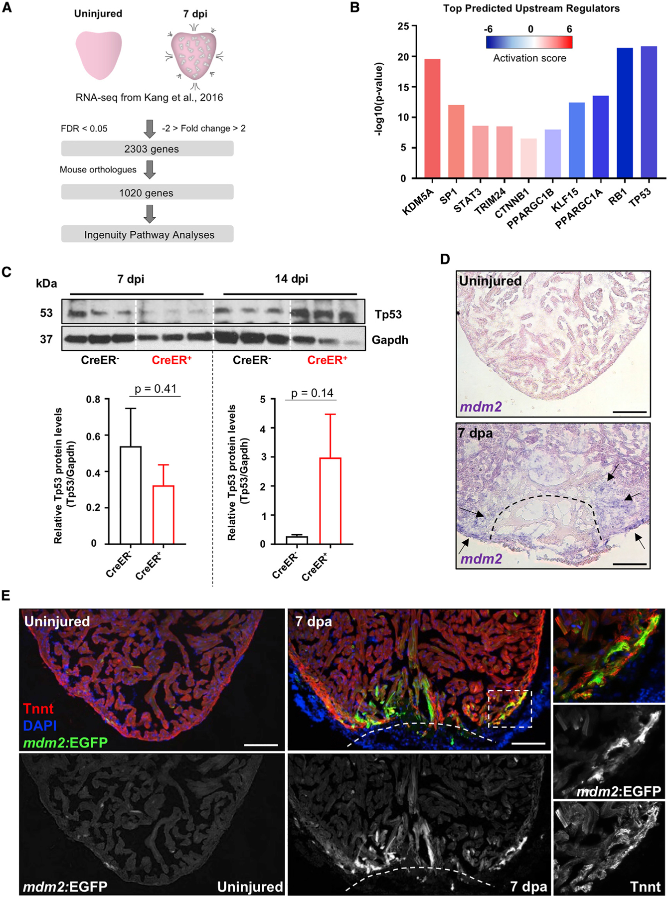
Figure 1. Zebrafish Tp53 and mdm2 Expression Are Dynamic upon Heart Injury.

(A) Experimental design and bioinformatical data analysis.
(B) Selected top transcription factors acting as upstream regulators of differentially expressed genes in adult hearts 7 days after initiation (dpi) of CM ablation.
(C) Western blot on protein extract of control (β-actin2:loxp-mCherry-STOP-loxp-DTApd36), 7 dpi and 14 dpi (cmlc2:CreER; β-actin2:loxp-mCherry-STOP-loxp-DTApd36) ventricles, with quantification of Tp53 protein. n = 8–10 pooled hearts per sample. Data show mean ± SEM (unpaired t test).
(D) ISH for mdm2 expression (violet) in sections of uninjured and 7 days after resection (dpa) ventricles. Arrows identify sites of increased mdm2 expression following ventricle amputation.
(E) Section images of uninjured and 7 dpa mdm2:EGFP hearts. “Tnnt” marks CMs. Boxes correspond to the region magnified in the right panels. Scale bars: 100 μm.
