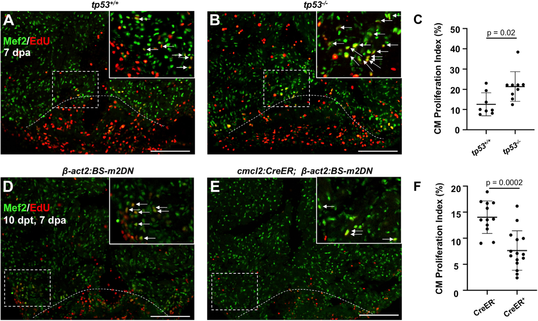
Figure 2. Tp53 or Mdm2 Modulation Alters CM Proliferation during Heart Regeneration.

(A and B) Images of tp53+/+ and tp53−/− hearts at 7 days post ventricular apex (dpa) resection. Arrows indicate Mef2+/EdU+ nuclei.
(C) Quantification of CM proliferation at 7 dpa indicating an increased proliferation index in tp53−/− mutants. n = 8 and 9 for tp53+/+ and tp53−/−, respectively.
(D and E) Images of β-act2:BS-m2DN and cmlc2:CreER; β-act2:BS-m2DN hearts at 7 dpa and 10 days post tamoxifen (dpt) administration. Arrows indicate Mef2+/PCNA+ nuclei.
(F) Quantification of CM proliferation showing a reduction in the proliferation index animals expressing a dominant-negative Mdm2. n = 12 and n = 15 for CreER− and CreER+, respectively. Scale bars: 100 μm. Data show mean ± SEM (Mann-Whitney U test).
