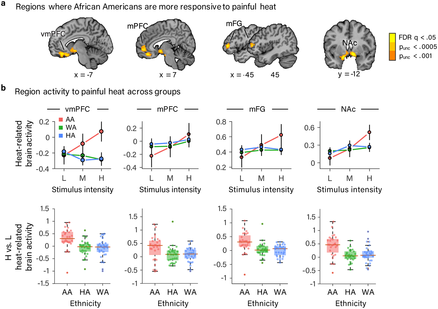
Figure 3.

Results of whole-brain voxel-wise GLM analysis showing brain regions exhibiting a stronger dose-response effect of painful heat in AA participants. (a) vmPFC, mPFC, bilateral mFG and bilateral NAc exhibited a steeper dose-response effect of painful stimulus intensity in AA compared to HA and WA participants (FDR corrected q < .05 (p < .000047)). For the purposes of display, we included voxels meeting two additional, more liberal, uncorrected voxel-wise thresholds (p < .0005 and p < .001) that were in contact with voxels meeting the more stringent FDR corrected threshold. (b) Data from the four regions (defined at p < .001, uncorrected and combining across hemispheres for bilaterally activated NAc and mFG). Top row: line plots depicting the mean parameter estimate for each level of stimulus intensity in each ethnic group plotted with error bars representing the within-subject SEM. In bottom row are box plots of the mean parameter estimate difference between high and low stimulus intensity in each ethnic group. Box plot elements: red center line, median; box limits, upper and lower quartiles; whiskers, limits of non-outlier data; points matched to box shading, subject means; dark red points, outliers. (a-b) Data are from 88 participants.
