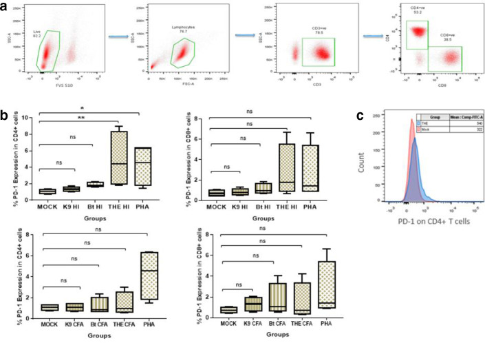
Fig. 1.
PD-1 expression upon exposure to HI bacteria. Peripheral blood mononuclear cells from healthy controls were stimulated in vitro with HI whole bacteria at an m.o.i. f 1 : 10 from the strains THE, B. thailandensis and K96243. (a) Gating strategy used for identifying T cell subsets. All gates were set using appropriate isotype controls. (b) PD-1 expression in CD4+ and CD8+ T cells following 18 h incubation with HI bacteria and culture filtrate antigen. Statistical analysis was performed using the Kruskal–Wallis test. P*<0.0125, P**<0.0025, P***<0.00025 with four Bonferroni comparisons. (c) An overlay histogram plot comparing mean fluorescence intensities of PD-1 in HI THE exposed and antigen-unexposed mock PBMCs. The data presented are representative of four individual experiments (n=4).

