
Figure 4.
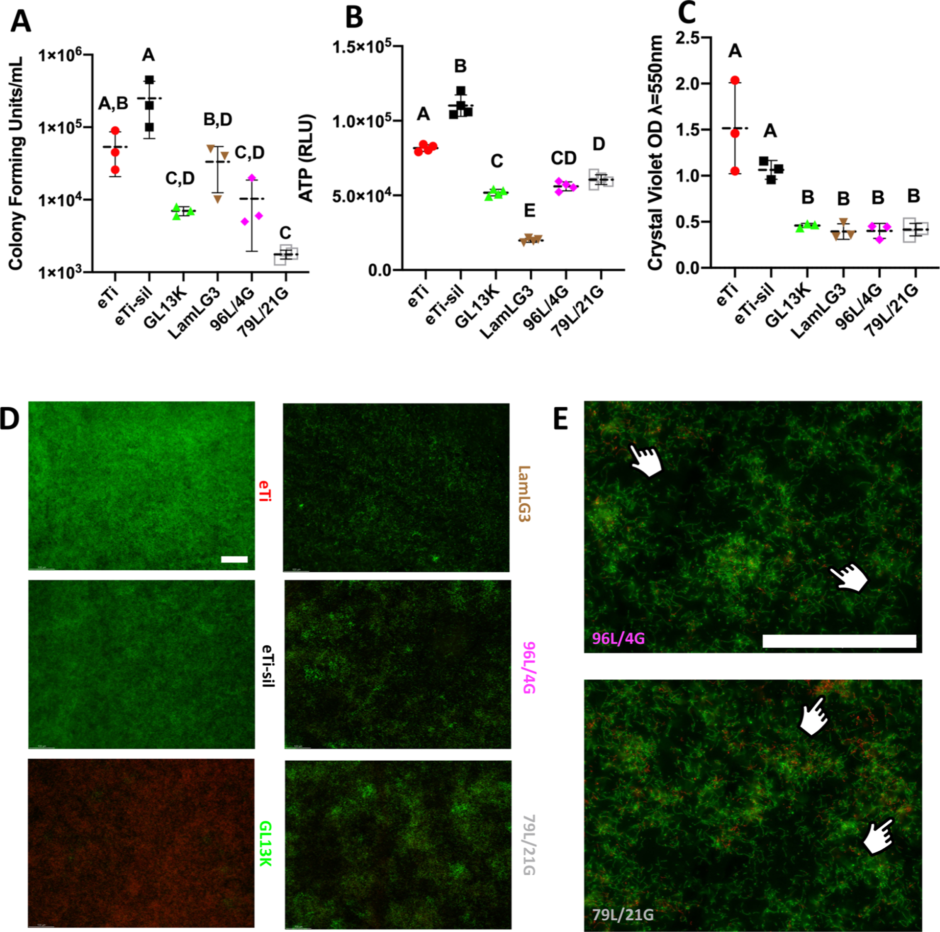
Antimicrobial activity of peptide coatings and controls against S. gordonii. (A) Colony Forming Units (CFUs) per mL. (B) ATP bioluminescence and (C) Crystal violet biomass of S. gordonii. (D) LIVE/DEAD micrographs of S. gordonii at ×10. (E) LIVE/DEAD micrographs at ×40. Fingers denote localized area of red (nonvital) staining. Scale bar is 100 μm for both magnifications. A p-value of <0.05 was considered statistically significant.