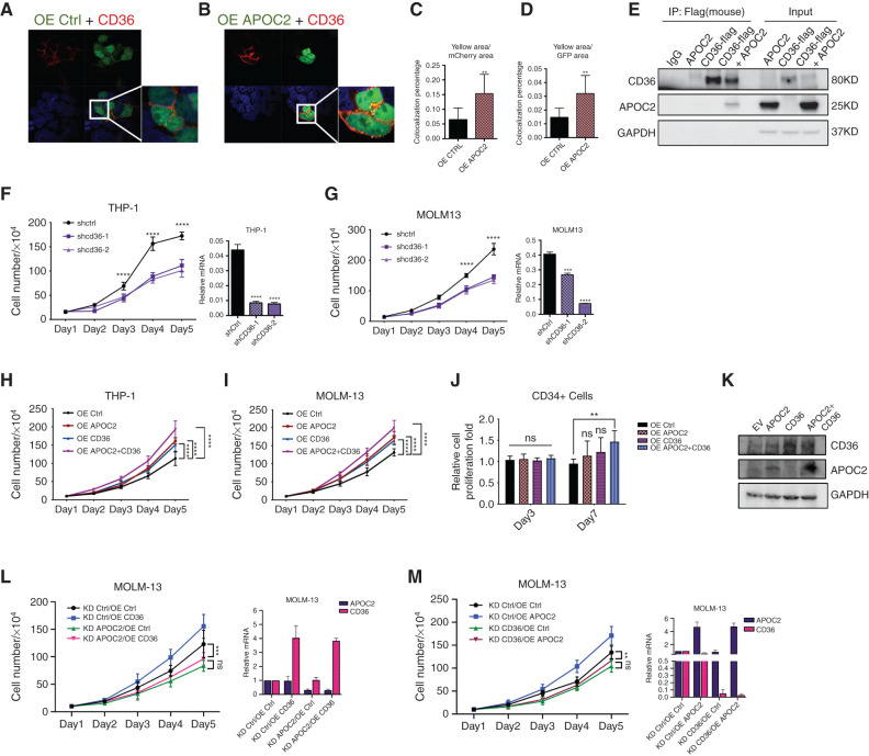
Figure 3.
APOC2 interacts and cooperates with CD36 to promote leukemia growth. A and B, Confocal microscopy images showing the colocalization of CD36 and APOC2. 293T cells were cotransfected with mCherry-CD36 and empty-GFP or mCherry-CD36 and APOC2-GFP plasmids and imaged 48 hours later by confocal microscopy. C and D, Quantitative analyses of colocalization area conducted by ImageJ. Pictures were taken from ten different fields for each slide (duplicate slides were used for each condition) and combined results of two different experiments were reported. E, Immunoprecipitation (IP) assay demonstrating the interaction between flag-CD36 and APOC2. 293T cells were cotransfected with flag-CD36 and APOC2 plasmids; flag antibody was used to pull-down CD36; APOC2 antibody was used to detect APOC2 in IP lysates as well as input in Western blot. F and G, Effects of tetracycline-controlled CD36 knockdown on cell proliferation in THP-1 and MOLM-13 cells. H and I, Effects of ectopic expression of APOC2, CD36, and both on cell proliferation in THP-1 and MOLM-13 cells. J, Viability of CD34+ cells ectopically expressing empty vector, APOC2, CD36, and both on days 3 and 7. K, The expression level of APOC2 and CD36 was detected by Western blot. L and M, Effects of knocking down APOC2 and CD36 on the proliferation of OE APOC2 and CD36 stable cells, respectively. mRNA levels of APOC2 and CD36 changes were confirmed by qPCR. The difference between groups was analyzed by unpaired t test (****, P < 0.0001; ***, P < 0.001; **, P < 0.01; *, P < 0.05; ns, not significant).

