
Figure 4.
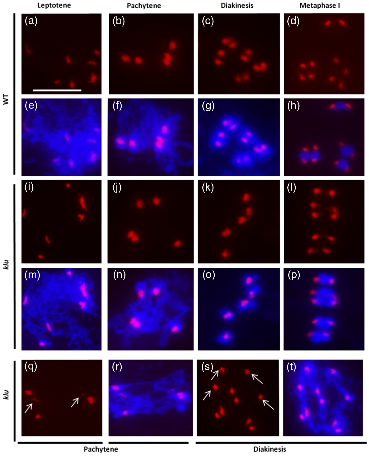
Defective centromere pairing during meiosis I in klu female meiocytes.
(a–d, i–l, q, s) Epifluorescence microscopy images of female meiotic spreads subjected to FISH analysis using a centromere-specific probe (red signal).
(e–h, m–p, r, t) Merged epifluorescence microscopy images of centromere (red) and 4′,6-diamidino-2-phenylindole (DAPI)-stained chromosomes (blue) in female meiotic spreads. (a, e), (b, f), (c, g) and (d, h) Wild-type female meiocyte at leptotene, pachytene, diakinesis and metaphase I, respectively.
(i, m), (j, n), (k, o) and (l, p) Apparently normal centromere signal in klu female meiocytes at leptotene, pachytene, diakinesis and metaphase I, respectively.
(q, r) klu female meiocyte at pachytene with unparied centromeres (white arrows).
(s, t) klu female meiocyte at diakinesis with a mixture of univalents (white arrows) and bivalents. Scale bars: 10 μm.