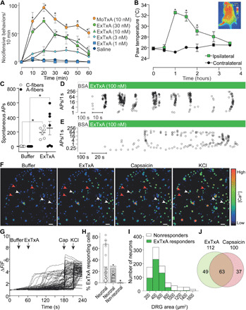
Fig. 3. Synthetic ExTxA and MoTxA activate sensory neurons in vivo and in vitro.

(A) Intraplantar injection of synthetic ExTxA and MoTxA caused dose-dependent nocifensive behaviors in C57BL/6 mice as well as (B) local hyperemia resulting in a significant increase in skin temperature. Inset: Thermal image illustrating the ExTxA-induced axon reflex vasodilation. *P < 0.05 by repeated-measures two-way ANOVA; n = 3 to 6. (C) Exposure of the cutaneous receptive fields of sensory neurons to synthetic ExTxA caused spontaneous AP firing in A-fibers (black circles) and C-fibers (open circles). (D and E) Representative recordings from the murine skin–saphenous nerve preparation illustrating ExTxA-induced activity in A-fibers (D) and C-fibers (E). Open circles, APs plotted as instantaneous frequency. (F to H) ExTxA (10 nM) induces Ca2+ influx in DRG neurons (white arrows), including capsaicin-sensitive nociceptors, but not nonexcitable cells (red arrowheads). (F) Pseudocolor image illustrating Ca2+ responses in DRG neurons (scale bar, 100 μm), and (G) corresponding traces from all neurons of one representative experiment. (H) Total percentage of neuronal cells (±1 μM TTX) and non-neuronal cells responding to ExTxA (10 nM). (I) Distribution of ExTxA-responding and nonresponding DRG neurons by cell size and (J) overlap with capsaicin-sensitive nociceptors. Data are presented as means ± SEM; *P < 0.05.
