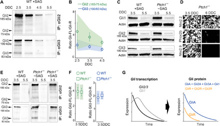
Fig. 2. Down-regulation of Gli genes establishes parallel temporal GliA/GliR gradients.

(A and B) Western blot of immunoprecipitated (IP) Gli2 or Gli3 protein from NSCs isolated at different DDC and quantification of protein ratios. (C) Western blot of Gli1-3 in WT and Ptch1−/− NSCs at 3.5DDC and 5.5DDC. (D) Immunofluorescence of Nkx2.2, Phox2b, and Nkx2.9 in Ptch1−/− ESC cultures at 3.5DDC and 6DDC. (E and F) Western blot of IP Gli2 or Gli3 protein from WT or Ptch1−/− NSCs isolated at 3.5DDC and 5.5DDC and differentiated in the presence or absence of SAG as indicated. Quantification of protein ratios of Gli2 and Gli3 bands in WT (+SAG) and Ptch1−/−(−SAG) NSCs at 3.5DDC. (G) Down-regulation of Gli genes produces parallel temporal GliA and GliR gradients. (B and F) Error bars, means ± SEM; shaded area, 95% confidence intervals (CIs).
