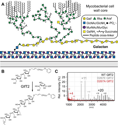
Fig. 1. The galactan and the mycobacterial cell envelope.

(A) Schematic depiction of the envelope of mycobacteria highlighting the mAGP (mycolyl-arabinogalactan-peptidoglycan). The glycans are depicted with the standard nomenclature. (B) Galactofuranosyl transferase 2 (GlfT2) extends the galactan to its final length. Polymerase activity can be probed in vitro using purified protein, synthetic acceptor, and sugar donor. (C) Overlayed matrix-assisted laser desorption/ionization–time-of-flight (MALDI-TOF) spectra produced by GlfT2 metal ion binding variants. Number of Galf residues added to acceptor noted above major product peaks.
