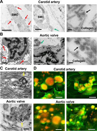
Fig. 1. Human cardiovascular tissue SMCs and VICs released EVs that aggregated and calcified in acellular ECM.

(A) SMCs in calcified human carotid artery and (B) VICs in calcified human aortic valve tissue transmission electron microscopy images (n = 5 donors, with representative images shown). Red arrows indicate EVs that likely budded from plasma membrane (scale bars, 500 nm), blue arrows indicate multivesicular bodies likely being released (scale bars, 500 nm), and black arrows indicate aggregated EVs in acellular collagen ECM (scale bars, 100 nm). (C) Transmission electron microscopy images of aggregated and calcifying EVs (yellow arrows indicate EVs with membrane hydroxyapatite formation) in collagen ECM in human carotid artery and aortic valve tissues (n = 5 donors, with representative images shown; scale bars, 200 nm). (D) Density-dependent scanning electron microscopy images of aggregated microcalcifications (yellow/orange color) in human carotid artery and aortic valve tissue ECM (green color); scale bars, 1 μm (n = 5 donors with two representative images shown).
