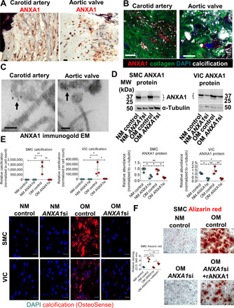
Fig. 4. ANXA1 knockdown attenuated human SMC and VIC calcification.

(A) ANXA1 immunohistochemistry (red color) in calcified (hematoxylin stain, purple color) human carotid artery and aortic valve tissues. Scale bars, 20 μm; n = 5 donors, with representative images shown and additional images included in fig. S5. (B) Human carotid artery and aortic valve tissue ANXA1 immunofluorescence (red color) near aggregated microcalcifications (OsteoSense 680, white color) in collagen ECM (CNA35-OG488, green color). Scale bars, 20 μm; n = 5 donors, with representative images shown. (C) Calcified human carotid artery and aortic valve tissue transmission electron microscopy (EM) of ANXA1 immunogold labeling (black dots) on EV in close proximity (arrow) in collagen ECM. Scale bars, 100 nm; n = 5 donors, with representative images shown, and additional images included in fig. S5. (D) Human SMC and VIC ANXA1 protein from cells cultured in control NM or OM and incubated with control siRNA (control) or ANXA1 siRNA (ANXA1si); quantification is the sum of both bands. Error bars are means ± SD from three donors. (E) Confocal microscopy images of microcalcifications (red color, OsteoSense 680 staining; blue color, DAPI) in SMCs and VICs cultured in NM or OM with control siRNA or ANXA1si; n = 3 donors, error bars are means ± SD; scale bars, 20 μm. (F) Alizarin red calcification stain and quantification for SMCs cultured in NM or OM with control, ANXA1si, or ANXA1si and recombinant human ANXA1 (rANXA1). Scale bars, 100 μm; error bars are means ± SD from three donors. ****P < 0.0001, **P < 0.01, and *P < 0.05, analyzed by analysis of variance (ANOVA).
