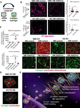
Fig. 6. ANXA1 inhibition suppressed EV microcalcification in 3D collagen hydrogels.

(A) Illustration showing 3D collagen hydrogel EV microcalcification experiment design. (B) Nanoparticle tracking analysis of single-EV quantity in conditioned media used for 3D collagen hydrogel EV experiments (n = 3 donors; analyzed by Welch’s t test). Confocal Z stack images and aggregate size quantification for DiR′-labeled (purple color) OM EV aggregates from SMCs [n = 3 donors with 352 control siRNA (control) and 225 ANXA1 siRNA (ANXA1si) EV aggregate areas quantified] and VICs (n = 3 donors with 65 control and 56 ANXA1si EV aggregate areas quantified). Scale bars, 20 μm; error bars are means ± SD; analyzed by Mann-Whitney test, ****P < 0.0001. (C) EV-generated microcalcifications (OsteoSense 680, red color) in 3D collagen hydrogels (CNA35-OG488, green color) using human SMC EV and VIC EV in conditioned NM or OM with control or ANXA1si; scale bars, 20 μm; n = 3 donors; error bars are means ± SD; ***P < 0.001 and *P < 0.05, analyzed by ANOVA. (D) Super-resolution microscopy images of OM conditioned media SMC EV and VIC EV calcification (OsteoSense 680) aggregates in 3D collagen hydrogels (CNA35-OG488). SMC EV x and y planes, 3200 nm; z, 800 nm; and VIC EV x, 10,800 nm, y, 9600 nm, z, 4800 nm; n = 3 donors, with representative images shown. (E) Working model in which altered calcium signaling may lead to increased calcium-binding proteins, including ANXA1 on EVs that promotes tethering of EVs trapped in ECM, leading to EV aggregation and formation and growth of microcalcifications.
